Home » Without Label » Shoulder Anatomy Diagram - Anatomy Of The Shoulder Ut Health San Antonio : These muscles form the outer shape of the shoulder and underarm.
Shoulder Anatomy Diagram - Anatomy Of The Shoulder Ut Health San Antonio : These muscles form the outer shape of the shoulder and underarm.
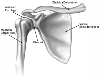
Shoulder Anatomy Diagram - Anatomy Of The Shoulder Ut Health San Antonio : These muscles form the outer shape of the shoulder and underarm.. Numerous muscles help stabilize the three joints of. The shoulder joint is the junction between the chest and the upper extremity. These muscles form the outer shape of the shoulder and underarm. The different types of connective tissues in the shoulder are bone, articular cartilage, ligaments, joint capsules, and bursa (see gross anatomy). The armpit and shoulder serve as the meeting place for the torso and arms, so major vessels close to the heart travel through these areas.
Elbow fractures icons orthopedic impingement body yoga anatomy back shoulder elbow fracture glenoid icons pain shoulder and elbow pain shoulder joint. #tcml #anatomy #charsi #shoulderjoint #diahram #mbbslike, comment, share, subscribefor any query tell me in comment section. 17 photos of the diagram of shoulder muscles and tendons. The muscles in the shoulder aid in a wide. Deltoides triangular refers to the front head of the.
Basic Anatomy Of The Shoulder Acro Physical Therapy Fitness from images.squarespace-cdn.com It is one of the most mobile joints in the human body, at the cost of joint stability. Numerous muscles help stabilize the three joints of. The shoulder joint (glenohumeral joint) is a ball and socket joint between the scapula and the humerus.it is the major joint connecting the upper limb to the trunk. Anatomy and injuries of the shoulder anatomical chart. The most flexible joint in the entire human body, our shoulder joint is formed by the union of the humerus, the scapula (or shoulder blade), and the clavicle (or collarbone). Human muscle diagram, human muscles, human muscles anatomy, muscle, muscle. Bones in shoulder, ligaments of the shoulder joint, parts of the shoulder joint, shoulder anatomy, shoulder joints and muscles, shoulder structure anatomy, shoulder tendon anatomy, shoulder tendons ligaments, human muscles, bones in shoulder, ligaments of the shoulder joint, parts of. The shoulder joint is formed where the humerus upper arm bone fits into the scapula shoulder blade like a ball and socket.
Sechrest, md narrates an animated tutorial on the basic anatomy of the shoulder.
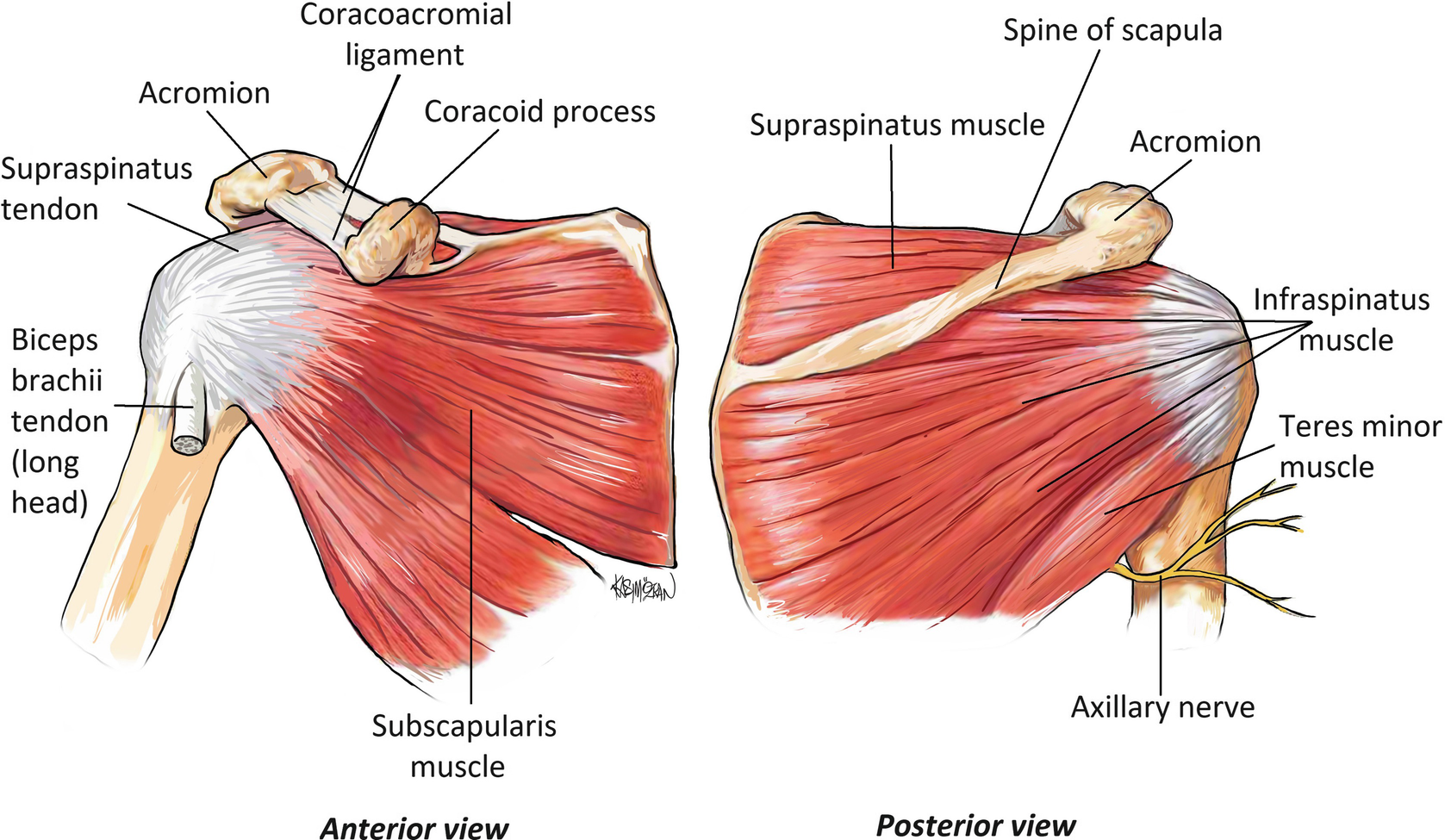
The most common symptoms of a torn shoulder labrum are: The muscles of the shoulder bridge the transitions from the torso into the head/neck area and into the upper extremities of the arms and hands. The shoulder plays a key role in the blood flow to the arms. Male shoulder ligaments and biceps muscles isolated in skeleton labeled chart on white labeled human anatomy diagram of male shoulder ligaments, connective tissue and biceps muscles isolated within the skeletal system frontal anterior view on a white background. The shoulder complex is composed of many different tissue types, and it is the connective tissue that provides the supportive framework for the shoulder's many functions. It causes pain in the area just outside the joint. The anterior shoulder pain usually develops when injury or inflammation occurs in the tendons that are attached to the shoulder joint. The shoulder is a very strong and flexible joint; The muscles in the shoulder aid in a wide. 17 photos of the diagram of shoulder muscles and tendons. However, more serious injuries, such as complete rotator cuff tears, may require surgical repair. Rotator cuff injuries are very common, affecting over 3 million people in the united states every year. The muscles of the shoulder support and produce the movements of the shoulder girdle.they attach the appendicular skeleton of the upper limb to the axial skeleton of the trunk.
This flexibility allows us to reach objects overhead as well as behind our backs. #tcml #anatomy #charsi #shoulderjoint #diahram #mbbslike, comment, share, subscribefor any query tell me in comment section. Find out in this anatomy of the shoulder quiz. The shoulder has about eight muscles that attach to the scapula, humerus, and clavicle. Pain in a man's body pain in a man's body on a gray background.
Shoulder Girdle Wikipedia from upload.wikimedia.org Sechrest, md narrates an animated tutorial on the basic anatomy of the shoulder. Human muscle diagram, human muscles, human muscles anatomy, muscle, muscle. Contents hide 1 anatomical terms. The primary function of the shoulder girdle is to give strength and range of motion to the arm. 2.1 bones of the shoulder girdle. Rotator cuff injuries are very common, affecting over 3 million people in the united states every year. The shoulder muscles consist of the deltoids and the rotator cuff group.the deltoids are the muscles that can be seen on the outside of the body, whilst the rotator cuff group is found within the shoulder joint itself, providing structural support and allowing the shoulder to perform many functions. Illustration of the shoulder anatomy and labrum.
Browse 3,096 anatomy of neck and shoulder stock photos and images available, or start a new search to explore more stock photos and images.
However, it requires considerable support from surrounding muscles and tendons. The shoulder has about eight muscles that attach to the scapula, humerus, and clavicle. Browse 3,096 anatomy of neck and shoulder stock photos and images available, or start a new search to explore more stock photos and images. 17 photos of the diagram of shoulder muscles and tendons. Name this muscle the largest of the shoulder group. #tcml #anatomy #charsi #shoulderjoint #diahram #mbbslike, comment, share, subscribefor any query tell me in comment section. 2.1 bones of the shoulder girdle. It causes pain in the area just outside the joint. The shoulder plays a key role in the blood flow to the arms. Four of them are found on the anterior aspect of the shoulder, whereas the rest are located on the shoulder's posterior aspect and in the back. The primary function of the shoulder girdle is to give strength and range of motion to the arm. Deltoides triangular refers to the front head of the. The most common symptoms of a torn shoulder labrum are:
The shoulder is a very strong and flexible joint; 17 photos of the diagram of shoulder muscles and tendons. Name this muscle that elevates the shoulder. Elbow fractures icons orthopedic impingement body yoga anatomy back shoulder elbow fracture glenoid icons pain shoulder and elbow pain shoulder joint. The anterior shoulder pain usually develops when injury or inflammation occurs in the tendons that are attached to the shoulder joint.
Shoulder Anatomy Springerlink from media.springernature.com Contents hide 1 anatomical terms. It causes pain in the area just outside the joint. 17 photos of the diagram of shoulder muscles and tendons. The most flexible joint in the entire human body, our shoulder joint is formed by the union of the humerus, the scapula (or shoulder blade), and the clavicle (or collarbone). All together they help hold your upper arm in place in the shoulder socket. Numerous muscles help stabilize the three joints of. Discuss tha agaonist/antagonist relationship of muscles. These symptoms may vary depending on the type of labral tear a person has.
All together they help hold your upper arm in place in the shoulder socket.
The muscles in the shoulder aid in a wide. Four muscles and their attached tendons make up the rotator cuff. #tcml #anatomy #charsi #shoulderjoint #diahram #mbbslike, comment, share, subscribefor any query tell me in comment section. The shoulder is a complex combination of bones and joints where many muscles act to provide the widest range of motion of any part of the body. All together they help hold your upper arm in place in the shoulder socket. Each of them aids in a specific motion of your shoulder. The most flexible joint in the entire human body, our shoulder joint is formed by the union of the humerus, the scapula (or shoulder blade), and the clavicle (or collarbone). The shoulder muscles consist of the deltoids and the rotator cuff group.the deltoids are the muscles that can be seen on the outside of the body, whilst the rotator cuff group is found within the shoulder joint itself, providing structural support and allowing the shoulder to perform many functions. What does a torn shoulder labrum feel like? The different types of connective tissues in the shoulder are bone, articular cartilage, ligaments, joint capsules, and bursa (see gross anatomy). Browse 3,096 anatomy of neck and shoulder stock photos and images available, or start a new search to explore more stock photos and images. 2.2 shoulder muscles and shoulder tendons. Discuss tha agaonist/antagonist relationship of muscles.