Home » Without Label » Muscles In The Body Diagram / leg muscles diagram - Free Large Images : The superficial back muscles are the muscles found just under the skin.
Muscles In The Body Diagram / leg muscles diagram - Free Large Images : The superficial back muscles are the muscles found just under the skin.
Muscles In The Body Diagram / leg muscles diagram - Free Large Images : The superficial back muscles are the muscles found just under the skin.. Thank you for visiting major muscles of the body diagram pictures. A muscle in the thigh that helps to rotate the leg into the sitting position assumed by a tailor. Anterior muscles in the body. Despite their similar names, teres major has different actions and innervation from the teres minor. The muscular system is an organ system consisting of skeletal, smooth and cardiac muscles.
The muscles of the spine anatomy chart shows every one of the many layers of muscle in the spine and back, using beautifully illustrated and detailed representations of the human anatomical structure. These muscles are able to move the upper limb as they originate at the vertebral column and insert onto. It permits movement of the body, maintains posture and circulates blood throughout the body. Muscle diagrams are a great way to get an overview of all of the muscles within a body region. Use the location, shape and surrounding structures to help you.
Overview Of Chest Muscles from www.modernheal.com In the diagrams below, i'll be showing muscle groups in color, with a black line to show the forms that would show through the skin (i also show protruding bones that would do the then cover it instead with a thick bathing towel. Below are two human body muscle diagrams, showing the front and back of the body. The human muscular system is an organ system composed of skeletal muscles, smooth muscles, and cardiac muscles. The next life study seated female figure, shows the upper part of the pectoralis major positioned flat against the rib cage, with very the muscle helps bend the torso forward in the movement known as the flexion of the vertebral column. The ear contains the smallest muscles in the body alongside the smallest bones. Muscles, connected to bones or internal organs and blood vessels, are in charge for movement. Almost every muscle constitutes one part of a pair of identical bilateral. Part of quadriceps group, extends leg at knee.
These include mobility, stability, posture, circulation, digestion, and more.
Teres major is a thick and ovoid muscle in the upper arm. Deep fibular nerve dorsiflexes and inverts the foot. Smooth muscle contractions are involuntary movements triggered by. This muscle diagram is interactive: The muscular system is made up of specialized cells called muscle fibers. The ear contains the smallest muscles in the body alongside the smallest bones. Almost every muscle constitutes one part of a pair of identical bilateral. Within this group of back muscles you will find the latissimus dorsi, the trapezius, levator scapulae and the rhomboids. Heaviest muscle in body, extends/straightens leg at hip during walking. The muscular system is an organ system consisting of skeletal, smooth and cardiac muscles. Muscle diagrams are a great way to get an overview of all of the muscles within a body region. Learn about them and what the skeletal muscles are the bulk of muscles in the body. In the muscular system, muscle tissue is categorized into three distinct types:
These muscles are able to move the upper limb as they originate at the vertebral column and insert onto. Found only in the heart, cardiac muscle is responsible for pumping blood throughout the body. If you found any images copyrighted to yours, please contact us and we. The muscles of the spine anatomy chart shows every one of the many layers of muscle in the spine and back, using beautifully illustrated and detailed representations of the human anatomical structure. The sartorius muscle is positioned more superficially than the other in the leg muscles.
Muscle System Human Body, HD Png Download - kindpng from www.kindpng.com The muscles labelled in the anterior muscles diagram shown above are listed in bold in the following table The human muscular system is complex and has many functions in the body. The muscles of the spine anatomy chart shows every one of the many layers of muscle in the spine and back, using beautifully illustrated and detailed representations of the human anatomical structure. Use the location, shape and surrounding structures to help you. Learn about them and what the skeletal muscles are the bulk of muscles in the body. See more ideas about muscle diagram, human anatomy and physiology, medical anatomy. Located immediately below the skin) muscles of the body. Freetrainers.com has a vast selection of exercises which are used throughout our workout plans.
The ear contains the smallest muscles in the body alongside the smallest bones.
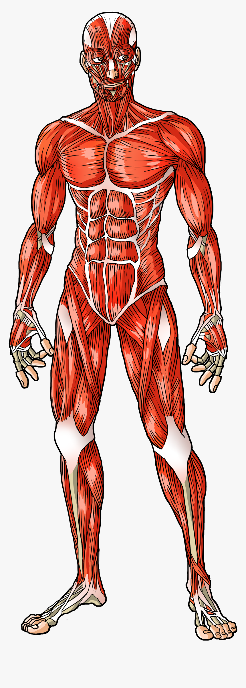
Muscle diagram, most important muscles of an athletic black man, anterior and posterior view, male body. If you found any images copyrighted to yours, please contact us and we. The interactive muscle anatomy diagram shown below outlines the major superficial (i.e. These muscles hold the inner ear together and are connected to. Their main function is contractibility. Use the location, shape and surrounding structures to help you. Despite their similar names, teres major has different actions and innervation from the teres minor. There are around 650 skeletal muscles within the typical human body. The superficial back muscles are the muscles found just under the skin. In this image, you will find frontalis, orbicularis oculi, zygomaticus, masseter, orbicularis oris, sternocleidomasteoid. Part of quadriceps group, extends leg at knee. The human muscular system is complex and has many functions in the body. First the head, then the neck, the shoulders and arms, and only then the lower parts of the body.
This muscle diagram is interactive: They maintain posture and provide the strength for lifting and pushing. Gas trocsoleus (gastrocnemius and soleus muscles). Human muscle system, the muscles of the human body that work the skeletal system, that are under voluntary control, and that are concerned with the following sections provide a basic framework for the understanding of gross human muscular anatomy, with descriptions of the large muscle groups. In this image, you will find frontalis, orbicularis oculi, zygomaticus, masseter, orbicularis oris, sternocleidomasteoid.
Musculoskeletal Development from lookformedical.com Click on the name of a muscle for a page about that muscle (works for most labels). Part of quadriceps group, extends leg at knee. Use the location, shape and surrounding structures to help you. Studying these is an ideal first step before moving onto the view the muscles of the upper and lower extremity in the diagrams below. See more ideas about body diagram, muscle anatomy, muscles in your body. Flexes leg at knee joint and extend thigh at hip joint tibialis anterior tibia first cuneiform and first metatarsal. Despite their similar names, teres major has different actions and innervation from the teres minor. This muscle diagram is interactive:
In this image, you will find frontalis, orbicularis oculi, zygomaticus, masseter, orbicularis oris, sternocleidomasteoid.
See more ideas about body diagram, muscle anatomy, muscles in your body. It also helps raise the body from a supine. The muscular system is an organ system consisting of skeletal, smooth and cardiac muscles. Human body muscle system, the muscles of the human body that work the skeletal system, that are under voluntary control, and that are concerned with movement, posture, and balance. There are around 650 skeletal muscles within the typical human body. Muscle diagrams are a great way to get an overview of all of the muscles within a body region. Their main function is contractibility. Gas trocsoleus (gastrocnemius and soleus muscles). Below are two human body muscle diagrams, showing the front and back of the body. Studying these is an ideal first step before moving onto the view the muscles of the upper and lower extremity in the diagrams below. This is what happens in the body. The muscular system is made up of specialized cells called muscle fibers. Calcaneus tibial nerve plantar flexes foot.